

Tinea Nigra
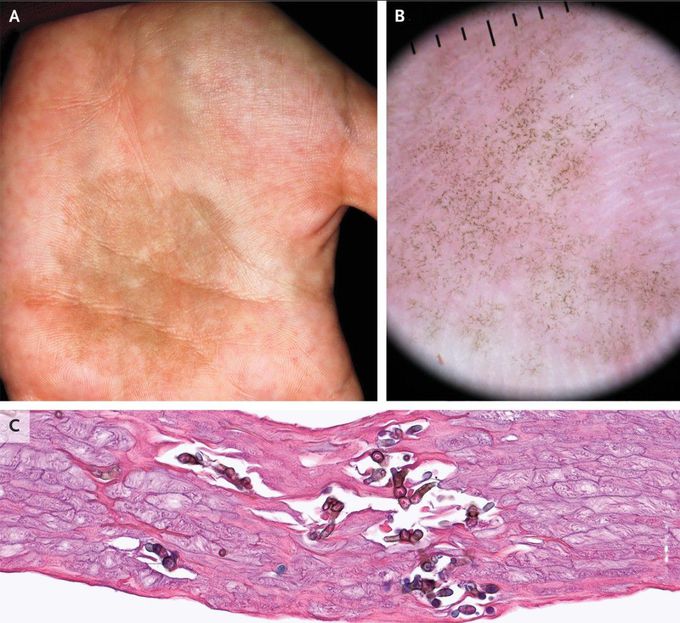
A 19-year-old woman presented to the dermatology clinic with a 6-month history of a slowly growing dark spot on her left palm. The lesion was asymptomatic and had not abated with soap-and-water rinses. The patient was a right-handed university student and reported no recent travel. Examination was notable for a nonscaling, nonpalpable brown patch on the left palm (Panel A). The right palm was not involved. Dermatoscopy revealed curvilinear brown strands — also known as pigmented spicules — that formed a reticular pattern distinct from the palmar furrows (Panel B; black lines indicate 1-mm intervals). Palmar skin scrapings that were prepared with potassium hydroxide and stained with hematoxylin and eosin showed pigmented yeast and hyphae (Panel C). A diagnosis of tinea nigra was made. Tinea nigra is a dematiaceous fungal infection typically seen in the tropical climates. It most commonly manifests as a hyperpigmented patch on the palm of the hand and may be mistaken for a melanocytic lesion, such as melanoma. After treatment with topical butenafine, the patient’s symptoms had abated at 3 months of follow-up.

