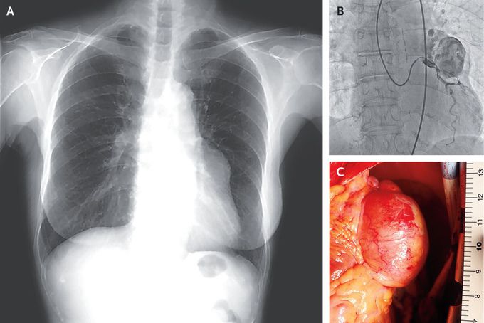


Giant Coronary-Artery Aneurysm
A 79-year-old woman with a history of stroke was referred to the cardiovascular outpatient clinic because of an abnormal radiographic finding along the left border of her heart (Panel A). The patient’s primary care physician had first noted the abnormality 6 years earlier on routine chest radiography. Owing to the patient’s absence of symptoms, the finding was monitored with serial radiographs, which showed that it had gradually increased in size. The cardiac examination was normal. Coronary computed tomography revealed an aneurysm measuring 45 mm in the greatest dimension arising from the left coronary artery — a finding that was consistent with a diagnosis of giant (measuring >20 mm in the greatest dimension) coronary-artery aneurysm. Subsequent cardiac catheterization showed blood flowing from the left coronary artery into the aneurysm and also into the pulmonary artery through a fistula (Panel B and video). Cardiac surgery involving ligation and plication of the aneurysm (Panel C) and closure of the fistula was performed. Giant coronary-artery aneurysms are often incidental and asymptomatic findings, but complications such as thrombosis, distal embolization, and rupture have been reported. The aneurysms are typically managed surgically, even in asymptomatic patients. The patient did well after surgery and was discharged 16 days later.
https://youtube.com/shorts/Q5QDgZynNjM?si=toiarnL66NpbMzhp

