MEDizzy
MEDizzy
DR.MOHAMMED IRFAN SHEIKH
DR.MOHAMMED IRFAN SHEIKHabout 1 year ago
Gallbladder Volvulus

Gallbladder Volvulus

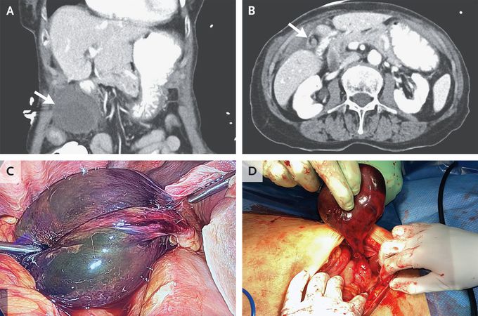
An 86-year-old woman presented to the emergency department with a 1-day history of nausea, vomiting, and pain on the right side of her abdomen. Her vital signs were normal. On physical examination, there was tenderness in the right upper quadrant, with a positive Murphy’s sign and involuntary guarding. The results of laboratory studies were normal. Computed tomography of the abdomen and pelvis revealed a distended gallbladder with a thickened wall outside the gallbladder fossa (Panel A, arrow). There was also swirling of the cystic artery and duct (Panel B, arrow) but no dilatation of the common bile duct. Gallbladder volvulus was suspected, and the patient was taken to the operating room. The diagnosis was confirmed on laparoscopy when a free-floating, necrotic gallbladder was observed encircling the cystic artery and duct (Panel C). The surgery was converted to an open procedure, and detorsion and resection of the gallbladder were successfully performed (Panel D). Gallbladder volvulus occurs when the gallbladder twists along the axis of the cystic artery and duct, compromising blood flow to the organ. Patients present with an acute abdomen, and the cause may be difficult to diagnose preoperatively. This patient did well after surgery and was discharged on postoperative day 2.

2
Other commentsSign in to post comments. You don't have an account? Sign up now!
Related posts
Flat Bones
Stomach
Foot X-ray AnatomyAnatomy of liverAbdominal aortic aneurysmCarcinoma gallbladder.Image of trachea blocked by chewing gum### Anatomy of the Heart: Summary The heart is a muscular organ responsible for pumping blood throughout the body, supplying oxygen and nutrients while removing waste products. It is located in the thoracic cavity, between the lungs, and is roughly the size of a fist. #### 1. **Structure:** - **Chambers:** The heart has four chambers: - **Right Atrium:** Receives deoxygenated blood from the body via the superior and inferior vena cavae. - **Right Ventricle:** Pumps deoxygenated blood to the lungs through the pulmonary arteries. - **Left Atrium:** Receives oxygenated blood from the lungs via the pulmonary veins. - **Left Ventricle:** Pumps oxygenated blood to the rest of the body through the aorta. - **Valves:** The heart contains four main valves that ensure unidirectional blood flow: - **Tricuspid Valve:** Located between the right atrium and right ventricle. - **Pulmonary Valve:** Located between the right ventricle and pulmonary arteries. - **Mitral (Bicuspid) Valve:** Located between the left atrium and left ventricle. - **Aortic Valve:** Located between the left ventricle and the aorta. #### 2. **Wall Layers:** - **Epicardium:** The outer layer, also known as the visceral pericardium. - **Myocardium:** The thick, muscular middle layer responsible for contraction. - **Endocardium:** The inner layer that lines the chambers and valves. #### 3. **Blood Supply:** - The heart receives its blood supply from the coronary arteries, which branch off the aorta. The **right coronary artery** supplies the right side of the heart, while the **left coronary artery** branches into the left anterior descending and circumflex arteries, supplying the left side. #### 4. **Electrical System:** - The heart's rhythmic contractions are controlled by its electrical conduction system, which includes: - **Sinoatrial (SA) Node:** The natural pacemaker located in the right atrium. - **Atrioventricular (AV) Node:** Receives impulses from the SA node and transmits them to the ventricles. - **Bundle of His and Purkinje Fibers:** Distribute the electrical impulse throughout the ventricles, leading to contraction. #### 5. **Function:** - The heart functions in two primary circulatory loops: - **Pulmonary Circulation:** Moves deoxygenated blood from the right side of the heart to the lungs for oxygenation. - **Systemic Circulation:** Distributes oxygenated blood from the left side of the heart to the rest of the body. The anatomy of the heart is crucial for its role in maintaining effective circulation and overall cardiovascular health. Understanding its structure helps in diagnosing and treating various heart conditions.

Recent MCQs


Show more MCQs

Recent flashcard sets


Show more flashcards