


Carcinoid Heart Disease
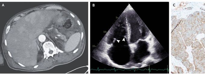
A 67-year-old man presented to the emergency department with a 1-month history of dyspnea and diarrhea and a 1-day history of abdominal pain. On physical examination, there was jugular venous distention with prominent V waves, a holosystolic murmur at the left lower sternal border, diffuse wheezing, and leg edema, but no flushing. Computed tomography of the abdomen showed thickening of the cecum and ascending colon, as well as numerous contrast-enhancing liver lesions (Panel A). Transthoracic echocardiography showed thickened, stiff tricuspid-valve leaflets with restricted mobility (Panel B [arrowheads] and Video 1) and severe tricuspid regurgitation (Video 2). The pulmonic valve was not clearly seen, but severe pulmonic regurgitation was noted. The 24-hour urinary 5-hydroxyindoleacetic acid level was 174 mg (reference value, <15). A biopsy of the liver mass showed a neuroendocrine tumor (Panel C, chromogranin A stain). A diagnosis of carcinoid syndrome with carcinoid heart disease in the context of a metastatic neuroendocrine tumor from a likely colonic primary site was made. Carcinoid heart disease occurs when high levels of vasoactive substances released from liver neuroendocrine metastases flow to the right heart and cause endocardial damage. Treatment with diuretics and octreotide was initiated, but the patient then opted for hospice care and died 1 month later.
