


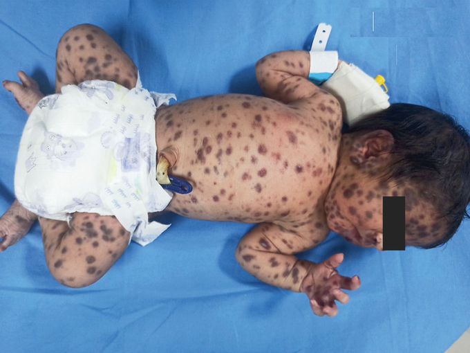
Congenital Langerhans-Cell Histiocytosis
A female infant delivered at term by cesarean section had a “blueberry muffin” rash at birth. On physical examination, multiple purple, papulonodular lesions measuring 5 to 10 mm in diameter were seen on her trunk, arms and legs, palms and soles, oral mucosa, and palpebral conjunctiva. There was no lymphadenopathy or hepatosplenomegaly, and the results of funduscopy were normal. The results of initial laboratory tests, including a complete blood count, a peripheral-blood smear, a reticulocyte count, a metabolic panel, and a coagulation test, were normal. A chest radiograph was normal, as were the results of ultrasonography of the head and abdomen. Neither rubella nor cytomegalovirus IgM or IgG was detected in the mother or the infant. A biopsy specimen of the skin obtained from the right thigh showed a dense infiltrate of histiocytes with grooved, kidney-shaped nuclei. Immunohistochemical staining of the specimen was positive for S100+ CD1a+ and negative for CD43 and myeloperoxidase, findings that confirmed a diagnosis of skin-limited congenital Langerhans-cell histiocytosis. Because of the lack of extracutaneous involvement, the lesions were expected to resolve without treatment. At follow-up 6 weeks after delivery, the skin lesions had resolved. The infant continues to be followed for signs of recurrence.
