MEDizzy
MEDizzy
DR.MOHAMMED IRFAN SHEIKH
DR.MOHAMMED IRFAN SHEIKHabout 1 year ago
Retinopathy in Malignant Hypertension

Retinopathy in Malignant Hypertension

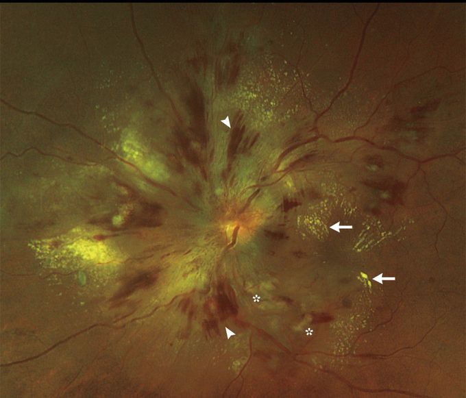
A 55-year-old man presented to the emergency department with a 1-week history of blurred vision in both eyes and headache. He had no chest pain, shortness of breath, or confusion. His blood pressure was 250/170 mm Hg. On ophthalmologic examination, the corrected visual acuity was 20/120 in the right eye and 20/80 in the left eye. Funduscopic examination revealed grade 4 hypertensive retinopathy, with disk edema in both eyes, flame-shaped hemorrhages (arrowheads), hard exudates (arrows), and cotton-wool spots (asterisks). Optical coherence tomography revealed foveal neurosensory detachment — a type of localized exudative retinal detachment — in both eyes, which explained the patient’s blurred vision. Laboratory studies revealed a blood urea nitrogen level of 41 mg per deciliter (15 mmol per liter; reference range, 8 to 26 mg per deciliter [3 to 9 mmol per liter]) and a creatinine level of 4.4 mg per deciliter (389 μmol per liter; reference range, 0.7 to 1.4 mg per deciliter [62 to 124 μmol per liter]), and urinalysis revealed hematuria and proteinuria. The patient was admitted to the hospital for management of malignant hypertension. While he was receiving medication for hypertension, his blood pressure improved; 5 months later, his visual acuity had improved to 20/30 in both eyes, and he had resolution of the foveal neurosensory detachments.

3
Other commentsSign in to post comments. You don't have an account? Sign up now!
Related posts
Flat BonesUveal Malignant Melanoma
Stomach
Foot X-ray AnatomyAnatomy of liverAbdominal aortic aneurysmImage of trachea blocked by chewing gum### Anatomy of the Heart: Summary The heart is a muscular organ responsible for pumping blood throughout the body, supplying oxygen and nutrients while removing waste products. It is located in the thoracic cavity, between the lungs, and is roughly the size of a fist. #### 1. **Structure:** - **Chambers:** The heart has four chambers: - **Right Atrium:** Receives deoxygenated blood from the body via the superior and inferior vena cavae. - **Right Ventricle:** Pumps deoxygenated blood to the lungs through the pulmonary arteries. - **Left Atrium:** Receives oxygenated blood from the lungs via the pulmonary veins. - **Left Ventricle:** Pumps oxygenated blood to the rest of the body through the aorta. - **Valves:** The heart contains four main valves that ensure unidirectional blood flow: - **Tricuspid Valve:** Located between the right atrium and right ventricle. - **Pulmonary Valve:** Located between the right ventricle and pulmonary arteries. - **Mitral (Bicuspid) Valve:** Located between the left atrium and left ventricle. - **Aortic Valve:** Located between the left ventricle and the aorta. #### 2. **Wall Layers:** - **Epicardium:** The outer layer, also known as the visceral pericardium. - **Myocardium:** The thick, muscular middle layer responsible for contraction. - **Endocardium:** The inner layer that lines the chambers and valves. #### 3. **Blood Supply:** - The heart receives its blood supply from the coronary arteries, which branch off the aorta. The **right coronary artery** supplies the right side of the heart, while the **left coronary artery** branches into the left anterior descending and circumflex arteries, supplying the left side. #### 4. **Electrical System:** - The heart's rhythmic contractions are controlled by its electrical conduction system, which includes: - **Sinoatrial (SA) Node:** The natural pacemaker located in the right atrium. - **Atrioventricular (AV) Node:** Receives impulses from the SA node and transmits them to the ventricles. - **Bundle of His and Purkinje Fibers:** Distribute the electrical impulse throughout the ventricles, leading to contraction. #### 5. **Function:** - The heart functions in two primary circulatory loops: - **Pulmonary Circulation:** Moves deoxygenated blood from the right side of the heart to the lungs for oxygenation. - **Systemic Circulation:** Distributes oxygenated blood from the left side of the heart to the rest of the body. The anatomy of the heart is crucial for its role in maintaining effective circulation and overall cardiovascular health. Understanding its structure helps in diagnosing and treating various heart conditions.

Recent MCQs


Show more MCQs

Recent flashcard sets


Show more flashcards