


Hampton’s Hump
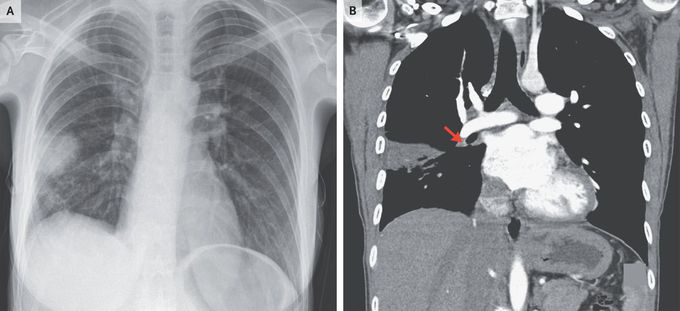
A 28-year-old woman presented to the emergency department with a 3-day history of dry cough and pleuritic chest pain. She had a history of ulcerative colitis and pulmonary embolism. Her heart rate was 87 beats per minute and her oxygen saturation 99% while she was breathing ambient air. On physical examination, fine crackles could be heard in the right lower lung. The legs were symmetric. Levels of troponin, d-dimer, and N-terminal pro–B-type natriuretic peptide were normal. An electrocardiogram showed deep S waves in lead I and T-wave inversions in lead III. A chest radiograph showed a dome-shaped, pleural-based opacity in the right lung (Panel A), consistent with Hampton’s hump, a finding that aroused concern about recurrent pulmonary embolism, even though her vital signs and results on laboratory testing were normal. Computed tomographic pulmonary angiography revealed filling defects in the right lower lobar pulmonary artery (Panel B, arrow) and its segmental branches as well as a peripheral wedge-shaped consolidation, confirming a diagnosis of pulmonary embolism with distal pulmonary infarction. Hampton’s hump, a radiographic sign of pulmonary infarction, is a rare finding in patients with pulmonary embolism. The patient started anticoagulation therapy, with a plan for lifelong therapy, and her symptoms resolved.
