


Pelvic Splenosis
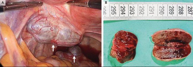
A 34-year-old woman presented to the hospital with a 2-year history of intermittent pelvic pain. Her surgical history included a cesarean section performed 10 years earlier and a partial splenectomy performed 29 years earlier after an automobile accident. The pelvic examination was notable for a soft parauterine mass that was mildly tender on palpation. Magnetic resonance imaging of the pelvis showed solid nodules with high T2-weighted signal intensity and restricted diffusion. An exploratory laparoscopy was performed and showed a hypervascular lesion (measuring 5 cm by 3 cm by 2.5 cm) on the left mesometrium and a second lesion (measuring 2.5 cm by 2 cm by 1.5 cm) on the mesorectum (Panel A, arrows), as well as a normal-appearing residual spleen in the left upper quadrant of the abdomen. Both masses were resected and appeared to be solid, encapsulated, and congested with blood (Panel B). Examination of a frozen section revealed splenic tissue. Splenosis is an uncommon condition in which splenic tissue autotransplants, typically after trauma or splenectomy. Pelvic splenosis can mimic gynecologic neoplasms. The patient’s pelvic pain resolved after the surgery.
