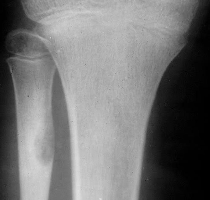


Dignosis of osteoid osteoma
Your healthcare provider will first ask about your symptoms and do a physical examination. They may ask you questions about the pain, such as: How long you’ve had pain. How severe the pain is. What helps to relieve the pain. Whether you’ve had an injury to the painful area. If your provider suspects an osteoid osteoma, they may suggest tests including: X-ray: This diagnostic imaging test creates pictures of your bone. In an X-ray, an osteoid osteoma will appear as thickened bone that surrounds a small central core. Three-phase bone scan: During a three-phase bone scan, your provider: Injects a radioactive material (radiotracer) into your vein. A camera detects this radiation and takes pictures of the radiotracer in your bone. The camera takes a picture of the blood that builds up in your bone and soft tissue. The camera takes another set of images of the same location two to three hours after the injection. This scan helps your provider find the exact location of the tumor. CT (computed tomography) scan or magnetic resonance imaging (MRI): A CT scan shows an image of your bone and can help identify an osteoid osteoma. An MRI is less accurate in showing an osteoid osteoma but can help rule out cancer. Biopsy: During a biopsy, your provider removes a sample of the tumor. They look at this tissue under a microscope for signs of an osteoid osteoma. This biopsy also helps them to rule out other conditions. Blood tests: Your provider may take blood tests to rule out an infection.
What is Rheumatoid Arthritis? | Johns Hopkins RheumatologyAnkylosing Spondylitis | HLA-B27, Pathophysiology, Signs & Symptoms, Diagnosis, TreatmentWhat is scoliosis?Osteosarcoma - Pathology, Symptoms, Diagnosis, TreatmentExternal nose bones

