MEDizzy
MEDizzy
alex chiurchi
alex chiurchiover 1 year ago
Myocarditis

Myocarditis

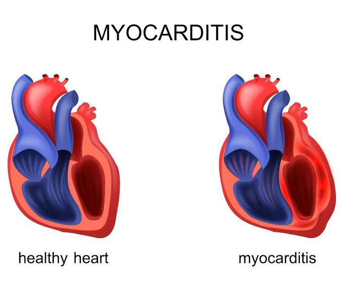
Myocarditis is a medical condition involving inflammation of the muscle tissue of the heart, known as the myocardium. This inflammation can damage the heart and affect its ability to effectively pump blood through the body. The causes of myocarditis can vary; it is often associated with viral infections, such as influenza virus, Epstein-Barr virus and cytomegalovirus. Other infections that can cause myocarditis include those caused by bacteria, fungi and parasites. However, it can also be caused by autoimmune diseases, allergic reactions to drugs or toxins, or be a complication of systemic diseases such as sarcoidosis. Symptoms of myocarditis can vary from mild to severe and may include fatigue, weakness, chest pain, dyspnoea (difficulty breathing), palpitations and swelling of the legs, ankles and feet. In some cases it may be asymptomatic and only be discovered during a routine medical examination or following complications such as heart failure or arrhythmias. The diagnosis of myocarditis can be complex, as the symptoms may be similar to other heart diseases. The physician may use a combination of examinations and tests, including electrocardiogram (ECG), echocardiogram, blood tests to detect markers of inflammation, and endomyocardial biopsy (taking a tissue sample from the heart for analysis). The treatment of myocarditis depends on the underlying cause and the severity of the symptoms. In many cases, rest and taking anti-inflammatory drugs may be sufficient to manage the inflammation and relieve symptoms, while in more severe cases, heart medications, such as beta-blockers or diuretics, may be prescribed to improve heart function. In more extreme cases, hospitalisation and the use of advanced therapies such as ventricular assist devices or heart transplantation may be required. It is important to emphasise that myocarditis can have long-term consequences for heart health and in some cases lead to permanent damage to the myocardium and increase the risk of arrhythmias, heart failure and sudden death.

7
Other commentsSign in to post comments. You don't have an account? Sign up now!
Related posts
Crohns diseaseCrohn’s disease of the ileumAlcoholic liver diseaseCirrhotic CardiomyopathyCirrhotic CardiomyopathyHeart and Lung Auscultation### Anatomy of the Heart: Summary The heart is a muscular organ responsible for pumping blood throughout the body, supplying oxygen and nutrients while removing waste products. It is located in the thoracic cavity, between the lungs, and is roughly the size of a fist. #### 1. **Structure:** - **Chambers:** The heart has four chambers: - **Right Atrium:** Receives deoxygenated blood from the body via the superior and inferior vena cavae. - **Right Ventricle:** Pumps deoxygenated blood to the lungs through the pulmonary arteries. - **Left Atrium:** Receives oxygenated blood from the lungs via the pulmonary veins. - **Left Ventricle:** Pumps oxygenated blood to the rest of the body through the aorta. - **Valves:** The heart contains four main valves that ensure unidirectional blood flow: - **Tricuspid Valve:** Located between the right atrium and right ventricle. - **Pulmonary Valve:** Located between the right ventricle and pulmonary arteries. - **Mitral (Bicuspid) Valve:** Located between the left atrium and left ventricle. - **Aortic Valve:** Located between the left ventricle and the aorta. #### 2. **Wall Layers:** - **Epicardium:** The outer layer, also known as the visceral pericardium. - **Myocardium:** The thick, muscular middle layer responsible for contraction. - **Endocardium:** The inner layer that lines the chambers and valves. #### 3. **Blood Supply:** - The heart receives its blood supply from the coronary arteries, which branch off the aorta. The **right coronary artery** supplies the right side of the heart, while the **left coronary artery** branches into the left anterior descending and circumflex arteries, supplying the left side. #### 4. **Electrical System:** - The heart's rhythmic contractions are controlled by its electrical conduction system, which includes: - **Sinoatrial (SA) Node:** The natural pacemaker located in the right atrium. - **Atrioventricular (AV) Node:** Receives impulses from the SA node and transmits them to the ventricles. - **Bundle of His and Purkinje Fibers:** Distribute the electrical impulse throughout the ventricles, leading to contraction. #### 5. **Function:** - The heart functions in two primary circulatory loops: - **Pulmonary Circulation:** Moves deoxygenated blood from the right side of the heart to the lungs for oxygenation. - **Systemic Circulation:** Distributes oxygenated blood from the left side of the heart to the rest of the body. The anatomy of the heart is crucial for its role in maintaining effective circulation and overall cardiovascular health. Understanding its structure helps in diagnosing and treating various heart conditions.Polycystic kidney diseaseAdult (autosomal dominant) polycystic kidney disease (ADPKD).Minimal change disease

Recent MCQs


Show more MCQs

Recent flashcard sets


Show more flashcards