


READING LUMBAR SPINE MRI
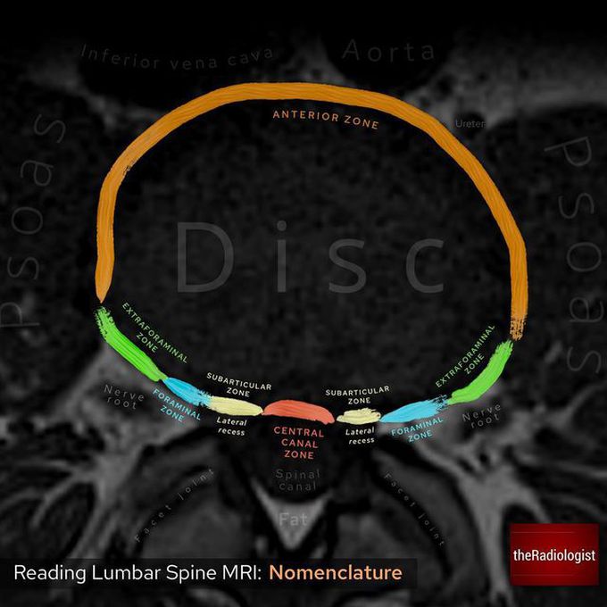
👨🏽💻Here we have an annotated diagram looking at the terms which help us describe the location of disc hernia rooms and osteophytes within the lumbar spine 👨🏽💻Let’s look at the zones in more detail: ▫️CENTRAL CANAL ZONE The posterior longitudinal ligament is especially thick in this region and so discs usually herniate slightly to the right or left rather than straight down the middle. These are then best described as ‘left central’ or ‘right central’ ▫️SUBARTICULAR (LATERAL RECESS) The most common region for disc herniation - this slice is taken at L4-5 meaning if the disc herniates into the subarticular zone, the L5 nerve root may be affected ▫️FORAMINAL ZONE Disc herniation is much less common here but can cause significant symptoms. If this occurs at L4-5 the L4 exiting nerve root would be affected ▫️EXTRAFORAMINAL ZONE Again not very common but more commonly missed compared with lateral recess herniation as it can be more difficult to see! Make sure to follow the exiting nerve root within the high signal fat and see high signal fat surrounding it 🔻Remember >80% of disc herniations occur at L4-5 and L5-S1 but more superior levels are affected with increasing age ▪️ Reference: ‘Lumbar disc nomenclature: version 2.0 Recommendations of the combined task forces of the North American Spine Society, the American Society of Spine Radiology and the American Society of Neuroradiology’ Published in the Spine Journal, 2014
Source: https://www.instagram.com/p/Co7Y7szNAZo/?igshid=NDk5N2NlZjQ=