


MV
MEDizzy Videosabout 9 years ago
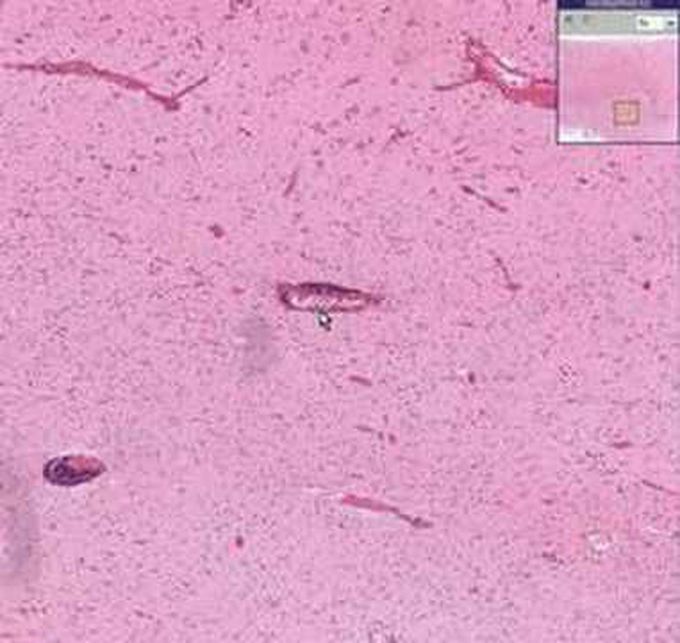
Viral encephalitis: Histopathology
In the attached video you can learn about the histopathological findings that can be typically seen in digital microscopic slide of a viral encephalitis brain specimen, which include some specific vascular findings. Timeline: 0:00 - Normal brain tissue microscopic morphology 0:43 - Vascular findings (vascular congestion and cuffing) in the brain specimen
Other commentsSign in to post comments. You don't have an account? Sign up now!
