


MV
MEDizzy Videosover 8 years ago
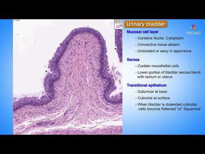
Urinary Bladder Histology: Histological Slide
In this short video you can study the stained section of the urinary bladder with all the relevant labellings and relations of different layers. Timeline: 0:00 - Introduction 0:13 - Mucosal cell layer 0:35 - Other parts of urinary bladder wall 0:51 - Serosa 1:22 - Artery, vein and connective tissue 1:34 - Spindle cells 1:49 - Transitional epithelium 2:50 - Lamina propria 3:07 - Detrusor muscle
Other commentsSign in to post comments. You don't have an account? Sign up now!
