


Orbital Rhabdomyosarcoma
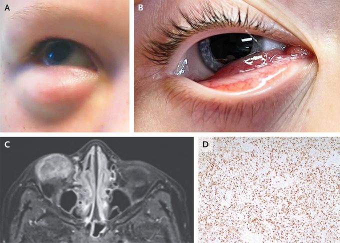
A previously healthy 8-year-old boy was brought to the emergency department with a 2-week history of painless swelling of the right lower eyelid. On physical examination, the swelling was found to be present during primary gaze (Panel A) but reduced during downward gaze (see video). A fixed mass measuring 1.5 cm by 2.5 cm was palpable within the eyelid. When the right eye was opened, erythematous subconjunctival tissue was seen protruding from under the lower eyelid (Panel B). Visual acuity and extraocular movements were normal in both eyes. Magnetic resonance imaging of the orbits showed a heterogeneous, circumscribed, T1-hypointense, T2-hyperintense mass along the anteroinferior portion of the right globe (Panel C, T1 fat-suppression sequence with contrast medium). An incisional biopsy was performed, and histopathological examination showed dense sheets of small, round, blue cells that stained positive for myogenin (Panel D). A diagnosis of embryonal rhabdomyosarcoma — the most common primary malignant orbital tumor in children — was made. A staging evaluation indicated stage 1 involvement, and the patient underwent mass debulking, adjuvant chemotherapy, and radiotherapy. At follow-up 3 months later, imaging showed no evidence of disease and the patient’s vision remained normal. Eyelid swelling that varies with eye movements should arouse suspicion for an extraocular muscle condition, including malignant tumors, as in this case. Erich J. Berg, D.O. Jeremy D. Clark, M.D. University of Louisville, Louisville, KY source: nejm.org

Living with HIV was one of the hardest experiences of my life. The fatigue, the emotional toll, and the uncertainty about the future weighed on me every single day. I had tried many treatments and medications, but nothing seemed to restore my health or energy the way I hoped.Out of both hope and desperation, I came across NaturePath Herbal Clinic. At first, I was skeptical but something about their natural approach and the powerful stories I read gave me the courage to try one more time.I began their herbal treatment program, and within a few weeks, I noticed small but meaningful changes more energy, better sleep, and a stronger immune system. Over the months, those improvements only grew. Today, I can truly say my life has changed. I feel healthier, more balanced, and finally in control of my well-being again.This isn’t just a testimony it’s a heartfelt recommendation to anyone living with HIV or any chronic condition. Don’t give up hope. I’m so grateful I gave NaturePath Herbal Clinic a chance. Visit their website to learn more: www.naturepathherbalclinic.com Email: info@naturepathherbalclinic.com
Acute Conjunctivitis (Pink Eye) | Allergic, Bacterial, Viral | Symptoms, Diagnosis, TreatmentWhat causes eye freckles? Are they dangerous?Strabismus: Everything You Need To KnowFATTY DEPOSITS of CHOLESTEROL around EYES | How to get rid of it?-Dr.Rajdeep Mysore|Doctors' CircleGlaucomaApical cystectomyMandibular deviation following Hemimandibulectomy
