


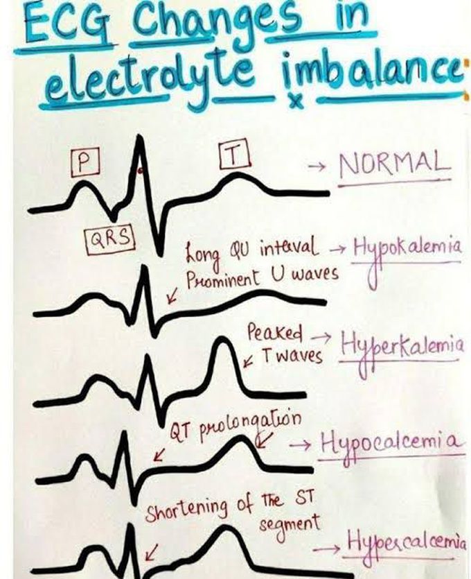
ECG changes in electrolyte imbalance
Hyperkalaemia: ECG changes All of the ECG changes that occur with a raised K+ concentration are non-specific and may affect any part of the ECG. The typical progressive changes of hyperkalaemia are as follows: Appearance of tall, pointed, narrow T waves. Decreased P wave amplitude, decreased R wave height, widening of QRS complexes, ST segment changes (elevation/depression), hemiblock (esp. left anterior) and 1st degree heart block. Advanced intraventricular block (very wide QRS with RBBB, LBBB, bi- or tri-fascicular blocks) and ventricular ectopics. Absent P waves, very broad, bizarre QRS complexes, AV block, VT, VF or ventricular asystole. ECG changes in decreasing order of frequency are: ST segment depression, decreased T wave amplitude, increased U wave height Cardiac arrhythmias Prolongation of the QRS duration, increased P wave amplitude and duration Various types of arrhythmias may occur in hypokalaemia. These may include atrial and ventricular ectopics, atrial tachycardia, heart blocks, VT and VF. Hypercalcaemia The main change is reduction in the Q-T interval on the ECG. The T wave duration is unaffected but the ST segment duration is shortened. Patients with hypercalcaemia have an increased sensitivity to digitalis and may present with a variety of arrhythmias. Hypocalcaemia The main ECG change is prolongation of the Q-T interval. There is no increase in T wave duration but the ST segment is prolonged. Magnesium It is useful to remember the following associations: In hypomagnesaemia, there is flattening of the T waves, ST segment depression, prominent U waves and, occasionally, a prolonged P-R interval occurs. In hypermagnesaemia, there may be a prolonged P-R interval and widened QRS complexes.
