


Mooren's ulcer
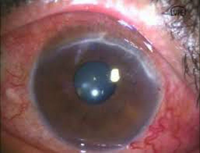
Mooren’s ulcer is characterized by painful peripheral corneal ulceration of unknown etiology. The disease generally begins with intense limbal inflammation and swelling in the episclera and conjunctiva [1]. Corneal changes begin within 2-3 mm from the limbus, first appearing as grey swellings that rapidly furrow, affecting the superficial one-third of the cornea and then proceeding circumferentially and centrally over 4-12 months.The bed of the furrow becomes vascularized, with vessels advancing into the base of the undermined edges of the ulcers These ulcers are often described as crescent-shaped and can leave behind an opaque and edematous central cornea. Alternatively, they can completely consume the corneal stroma, replacing it with a thin fibrovascular membrane.Inflammation is not seen in the sclera adjacent to the peripheral ulcers, nor does it affect the underlying Descemet’s membrane. Destruction of the cornea generally affects stromal tissue only, leaving behind an intact endothelium and epithelium.The central edges of the ulcer can develop an overhanging edge with or without opacification, and neovascularization of the cornea can occur, extending from the limbus into the ulcer bed.Neovascularization can occur up to the advancing edge of the ulcer but not beyond it.
