


A Ruptured Aorta- Chest X-ray
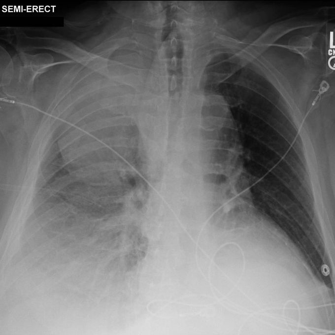
🎯CXR: Right pleural effusion. Large mass projects over the right upper chest. ⠀⠀⠀⠀⠀⠀⠀⠀⠀ 🎯CT: Intramural aortic haematoma extends from the aortic arch to just below the level of the right inferior pulmonary vein. Beginning at the level of the carina, there is blood outside the aorta anterior to the spine, indicating aortic rupture. Small bilateral pleural effusions, both contain high-density blood. The right chest mass seen on the plain film is extremely dense, indicating that it is blood. The mass has an "extrapleural fat sign", i.e. there is inward displacement of the extrapleural fat by the haematoma, indicating that the mass is located outside the pleura. ⠀⠀⠀⠀⠀⠀⠀⠀⠀ 🎯This case illustrates a ruptured aorta, with an intramural haematoma and bleeding into the soft tissues around the aorta, the pleural space and the right chest wall. ⠀⠀⠀⠀⠀⠀⠀⠀⠀ 👤Case courtesy of Stefan Tigges, Radiopaedia.org, rID: 97972
Source: https://www.instagram.com/p/CbH0zzRIZI9/?igshid=YmMyMTA2M2Y=