SM
Syed Mohammad Hasanover 2 years ago

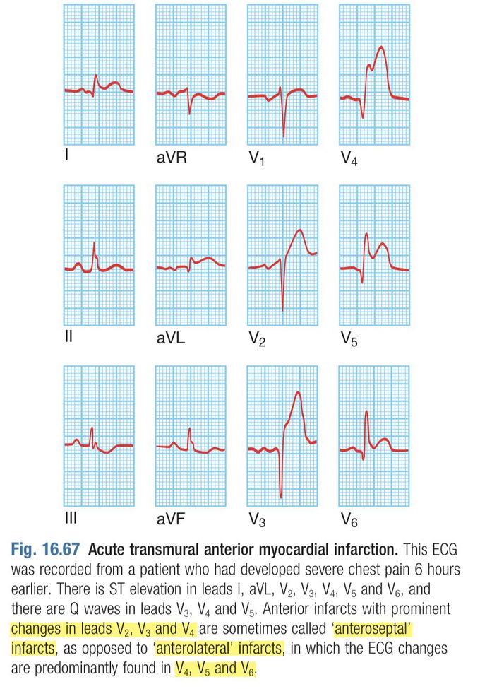
EKG Image of Acute Transmural Anterior MI
Anterior MI on the basis of EKG lead findings is sometimes distributed into anteroseptal infarcts and anterolateral infarcts. In anteroseptal infarcts, the EKG findings are usually seen in leads V2, V3, and V4 in contrast to anterolateral infarcts that are observed in leads V4, V5, and V6. The above-provided image gives an overview of the EKG picture of an extensive anterior wall MI. Image Source and Text Reference: Davidson’s Principles and Practice of Medicine, 23rd Edition, Page No: 497.
Other commentsSign in to post comments. You don't have an account? Sign up now!
Related posts
Supraventricular tachycardia- DiagnosisPatent Ductus Arteriosus- SymptomsAtrial Septal Defect- Diagnosis
https://www.facebook.com/AspenDoseCBDGummiesPage/Lipofit⢠Canada | Advanced Weight Loss Supporthttps://www.facebook.com/StableGripSafetyBarOfficial/Pulsar Vexline Review 2026 - Legit Or Scam Trading Platform? Fact CheckGelatine Sculpt | Official WebsiteBurnSlim Official | Support Your Weight Management Journey
https://www.facebook.com/AspenDoseCBDGummiesPage/Lipofit⢠Canada | Advanced Weight Loss Supporthttps://www.facebook.com/StableGripSafetyBarOfficial/Pulsar Vexline Review 2026 - Legit Or Scam Trading Platform? Fact CheckGelatine Sculpt | Official WebsiteBurnSlim Official | Support Your Weight Management Journey

