


Mohs surgery
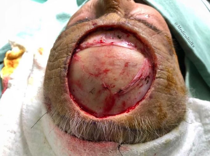
Mohs surgery for an extensive forehead squamous cell carcinoma!! Post-op pictures are coming next! Squamous cell carcinoma is a type of non-melanoma skin cancer that arise as a result of uncontrolled growth of the squamous cells in the epidermis layer of the skin and is suspected in patients with a rough, scaly nodule or non-healing, painless ulcer that develops in the setting of a scar or chronic inflammatory lesion. Risk factors are sun and radiation exposure, chronic wounds, burns, or scars. It is rapid growing and invasive, thus diagnosis is made by skin biopsy that includes the deep reticular dermis to assess the depth of the invasion. Small or low-risk lesions are managed with surgical excision or local destruction with cryotherapy or electrodessication, which the high risk and larger lesions should be referred for Mohs surgery. This patient underwent a Mohs micrographic surgery to treat his condition, which involves a sequential removal of thin skin layers with microscopic inspection to confirm that the margins have been cleared of malignant tissue. This technique has the highest cure rate for basal cell carcinomas and in smaller lesions it provides the least disruption to surrounding tissues, making it ideal for delicate areas such as the perioral region, nose, lips, ears and in this case, scalp. A graft surgery followed shortly after to cover the defect. If left untreated, squamous cell carcinoma can cause extensive local destruction and lymphatic or distant metastases. Photo by @dr.andremiolo
Hemodynamic stimuli&nonhemodynamic stimuliEffects of sugar on teeth
