
Abeer Fatimaabout 2 months ago

MRI Findings in Multiple Sclerosis
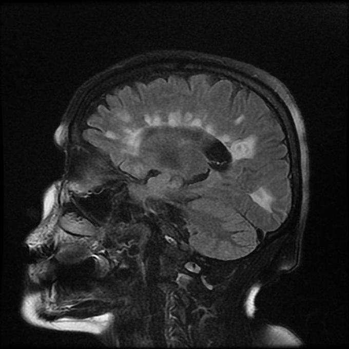
Many modalities of MRI are essential in diagnosis of multiple sclerosis. These sequences are discussed as follows: 1. T1 - Isointense to hypointense lesion - Thin corpus callosum - Hyperintense lesions when associated with brain atrophy 2. T2 - Hyperintense lesion - Edema surrounds the lesion 3. SWI - Central vein sign 4. FLAIR - Hyperintense lesions - Ependymal dot-dash sign - Dawson’s fingers 5. T1 C+ - Enhanced active lesions - Open ring sign 6. DWI/ADC - High or low ADC in active lesion 7. MR Spectroscopy - Reduced NAA peaks in plaques - Increased choline and lactate in acute phase Reference: https://radiopaedia.org/articles/multiple-sclerosis Image via: https://radiopaedia.org/articles/dawson-fingers
Other commentsSign in to post comments. You don't have an account? Sign up now!

