


Tedrik Markarian over 4 years ago
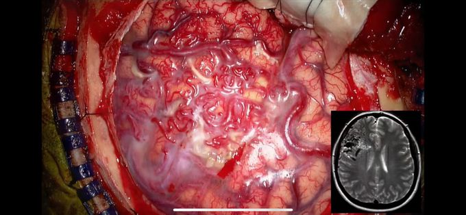
Cranial AVM (Arteriovenous malformation)!
An AVM is an abnormal pial vascular malformation. It contains a main nidus, but does not contain an intervening capillary bed. It can contain calcification, glial tissue, and blood. Furthermore, one of the main feeders can be part of the Circle of Willis (MCA, PCA, etc.), and draining veins can drain the AVM into a dural sinus (superior sagittal sinus, transverse sinus, sigmoid sinus, etc.). Treatment includes embolization using Onyx or surgical resection of the AVM. Patients diagnosed with an AVM are typically at risk for developing cranial hemorrhages if the lesion ruptures. Credits: Dr. Aaron Cohen-Gadol
Other commentsSign in to post comments. You don't have an account? Sign up now!
Related posts
