
Which explains the patients condition?
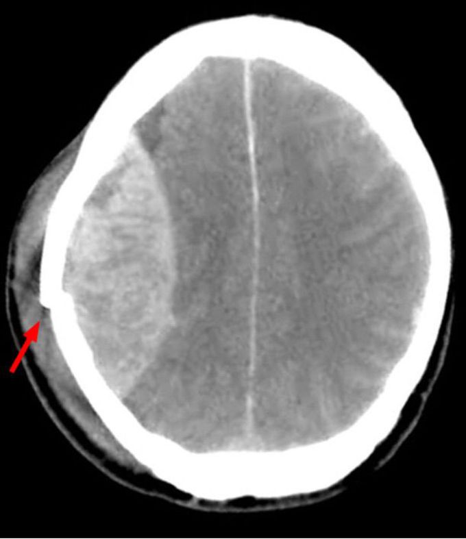
A 30-year-old male is brought to a trauma center by an emergency responder team after a vehicle accident. The patient was driving a motorcycle and collided with a bus. The EMT personnel found the patient alert with a short amnesia of the moments preceding the accident. In the ER, the patient is drowsy but still follows commands. Both his pupils are symmetrical and responsive to light with no other evident deficits. While the patient is being transferred from the computed tomography (CT) scanner room to observation floor, his level of consciousness declines to stupor, his left pupils became unresponsive to light, and his blood pressure rises to 190/110 mm Hg. His CT scan is given on the right. A.Tearing of the middle meningeal artery B. Dural arteriovenous fistula C. Tearing of bridging veins D. Charcot-Bouchard aneurysm E. Arteriovenous malformation
A, is an epidural hematoma, the temporal bone fracture is above the middle meningeal artery and the epidural hematoma looks like a lens on CT. The pacient worse because the epidural hematoma promote mass effect, with herniation of uncus, compressing the brainstem and lowering consciousness level. Without urgent surgery, the prognostic is bad. When the blood came from a vein rupture, the image shows blood above the brain, spread in the subdural space, and no circunscribed like this image. Charcot-bouchard aneurisms arn't seen in the CT, they are more likely to tease intracerebral bleeding, in the parenchima.

There are no displacement of meninge, is not epidural. Low mass efect.


Most likely A. If you read the description closely all the other options cancel out.