


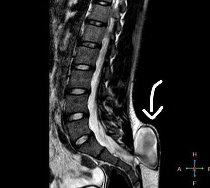
Spina Bifida (myelomeningocele)
MRI depicts a relatively large case of what was diagnosed to be myelomeningocele. Early on during the development of the fetus, the ectoderm (outmost later of a fertilized egg) begins to form a ridge which will later become the neural tube. However, in spina bifida patients, this neural tube does not close properly. As a result, usually in the lumbar or sacral regions, there is an absence of a vertebral arch (composed of the spinous process, transverse process, pedicles, lamina, and the articular process). There are different forms of spina bifida for which myelomeningocele is the most severe as the spinal cord and the surrounding meninges (dura, arachnoid, pia) protrudes out of the vertebral body. Myelomeningocele is usually seen in Arnold Chiari II Malformation for which findings include obstructive hydrocephalus, myelomeningocele, corpus callosum dysgenesis, no septum pellucidum, sometimes extended cerebellum tonsils into the foramen magnum, small posterior fossa, etc. It is known that a Vitamin B9 deficiency during the development of the fetus is a risk factor for spina bifida. Now, as for diagnosis of spina bifida, one must look for elevated alpha fetoprotein. Furthermore, as with ordered blood tests, one must look for estriol, inhibin A, and human chorionic gonadotropin. All these tests are usually accompanied with an ultrasound.
