


Mishal Shanalmost 2 years ago
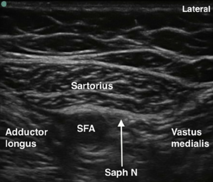
Ultrasound of the Adductor Canal
This image provides an ultrasound view of the adductor canal. The femoral artery and saphenous nerve can be visualized. At this site, an ultrasound-guided block of the saphenous nerve can be performed.
Other commentsSign in to post comments. You don't have an account? Sign up now!
