


Disseminated Fusariosis
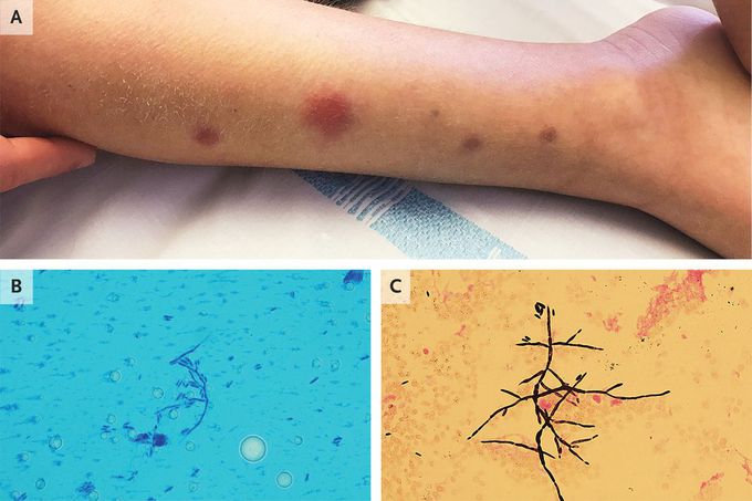
An 8-year-old boy presented with subcutaneous nodules associated with a 1-week history of fever 2 months after starting treatment for a relapsing B-cell leukemia. He had had pancytopenia for 57 days, and laboratory studies showed a hemoglobin level of 7.5 g per deciliter (reference range, 11.5 to 14.5), a white-cell count of 200 per cubic millimeter (reference range, 5000 to 11,000), and a platelet count of 12,000 per cubic millimeter (reference range, 150,000 to 400,000). Fevers persisted despite the initiation of broad-spectrum intravenous antibiotic and antifungal treatment, and 1 week later, the subcutaneous nodules developed. Physical examination revealed nodules ranging from 5 to 18 mm in diameter on the chest, back, arms, and legs (Panel A). A biopsy specimen of a nodule was obtained. Microscopic examination of a dry smear stained with lactophenol cotton blue revealed septated hyphae and spores (Panel B), and Fusarium dimerum was grown in culture. Gram’s staining of a blood culture revealed hyphae and spores (Panel C), and cultures also revealed F. dimerum. The patient completed a prolonged course of treatment with antifungal therapy. His fever resolved within 11 days, and the skin lesions resolved within 20 days.

Something you don't see everyday if his immune system was healthy
