


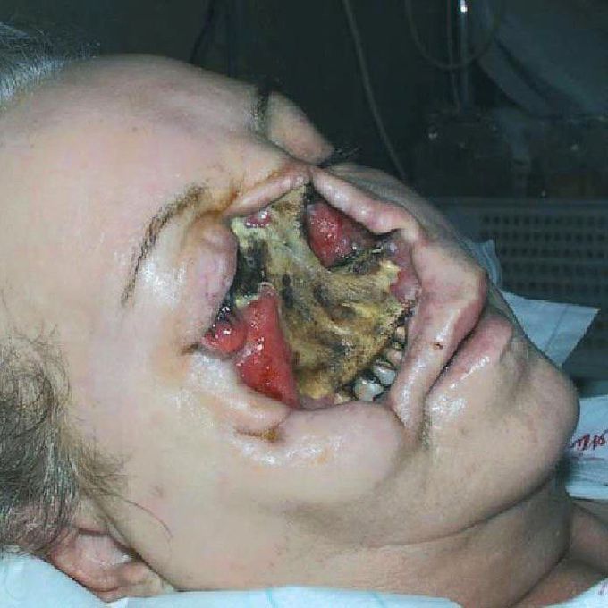
Combined Cranial Mucormycosis and Aspergillosis!
A 50-year-old woman was referred to with local symptoms of periodontitis. Three weeks earlier, she had undergone a right first premolar tooth extraction under local anesthesia without complications.Biopsy specimens were taken from the sides of the necrotic lesion and sent for histological examination. The outstanding microscopic features were of an intense, acute and chronic inflammatory infiltration, including scattered broad, branching, aseptate fungal hyphae, suggestive of infection by Mucoraceae Furthermore, a wound swab from the maxillary sinus was cultured evidencing conidia, associated with flower-like conidiophores, suggestive of Aspergillus. The diagnosis of combined mucormycosis and aspergillosis of the rhinocerebral region was made 6 weeks after hospital admission.
Was a there any information on why she waited so long, what reconstruction surgery was done, and was there any chronic issues as a result of this. Was it the extraction or post procedure that caused this.

Hemodynamic stimuli&nonhemodynamic stimuliEffects of sugar on teeth
