
Gross Anatomy of the Middle Ear - Boundaries ,Contents and Functions ( Animation )
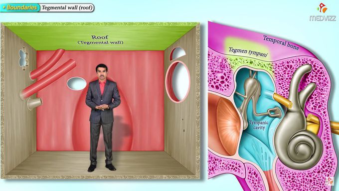
Follow On Instagram :- https://www.instagram.com/drgbhanuprakash Channel Memberships : https://www.youtube.com/channel/UCG5TBPANNSiKf1Dp-R5Dibg/join Gross anatomy of the Middle Ear - Boundaries, Contents and Functions (Animation) The ear can be split into three parts; external, middle and inner. The middle ear lies within the temporal bone, and extends from the tympanic membrane to the lateral wall of the inner ear. The main function of the middle ear is to transmit vibrations from the tympanic membrane to the inner ear via the auditory ossicles. Parts of the Middle Ear The middle ear can be divided into two parts: Tympanic cavity – located medially to the tympanic membrane. It contains three small bones known as the auditory ossicles: the malleus, incus and stapes. They transmit sound vibrations through the middle ear. Epitympanic recess – a space superior to the tympanic cavity, which lies next to the mastoid air cells. The malleus and incus partially extend upwards into the epitympanic recess. Borders The middle ear can be visualised as a rectangular box, with a roof and floor, medial and lateral walls and anterior and posterior walls. Roof – formed by a thin bone from the petrous part of the temporal bone. It separates the middle ear from the middle cranial fossa. Floor – known as the jugular wall, it consists of a thin layer of bone, which separates the middle ear from the internal jugular vein Lateral wall – made up of the tympanic membrane and the lateral wall of the epitympanic recess. Medial wall – formed by the lateral wall of the internal ear. It contains a prominent bulge, produced by the facial nerve as it travels nearby. Anterior wall – a thin bony plate with two openings; for the auditory tube and the tensor tympani muscle. It separates the middle ear from the internal carotid artery. Posterior wall (mastoid wall) – it consists of a bony partition between the tympanic cavity and the mastoid air cells. Superiorly, there is a hole in this partition, allowing the two areas to communicate. This hole is known as the aditus to the mastoid antrum. #middleearanatomyanimation #middleear #drgbhanuprakash #middleeargrossanatomy #anatomyofmiddleear #middleearboundaries #middleearcontents #middleearfunctions #middleeargrossanatomy #middleearvideo #middleearusmle #middleearexplained #middleearlecture #middleearanatomy #earanatomy #anatomyoftheear #middleearfunction #middleearborders #middleearanimation #anatomyoftheear #usmlestep1 #earanatomy