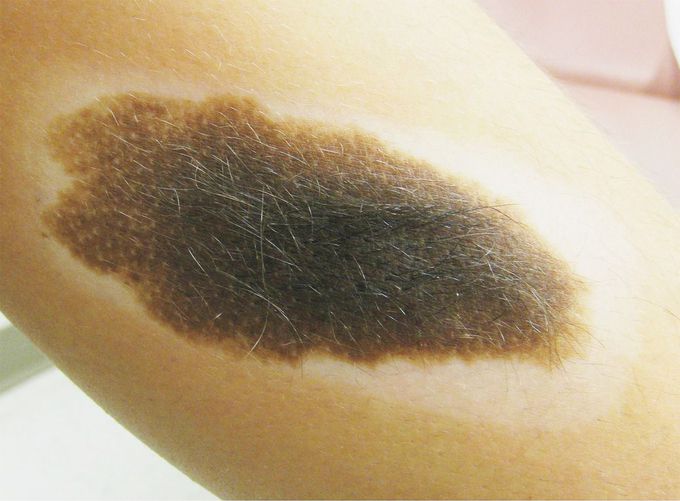


Halo Phenomenon
An otherwise healthy 14-year-old boy presented for evaluation of an 11.0-cm-by-4.5-cm congenital melanocytic nevus on the medial left shin that had been followed closely by the dermatology service since the boy was 3 years of age. The nevus, present since birth, had grown with the patient and had typical features, including variegated color, hypertrichosis, small scattered macules, and an irregularly scalloped border. In addition, a white halo surrounding its borders had gradually appeared during the past year. Halo nevi (also known as Sutton's nevi or leukoderma acquisitum centrifugum) are nevi that develop a border of depigmentation resembling a halo. The halo phenomenon is thought to involve destruction of melanocytes by CD8+ cytotoxic T lymphocytes and has been associated with vitiligo and rare spontaneous regression of melanocytic lesions. Because of the risk of malignant transformation inherent in a congenital melanocytic nevus, as well as the psychosocial distress associated with continued growth of the nevus, the patient is undergoing serial excision of the lesion. Gerhard S. Mundinger, M.D. Johns Hopkins Hospital, Baltimore, MD source: nejm.org

