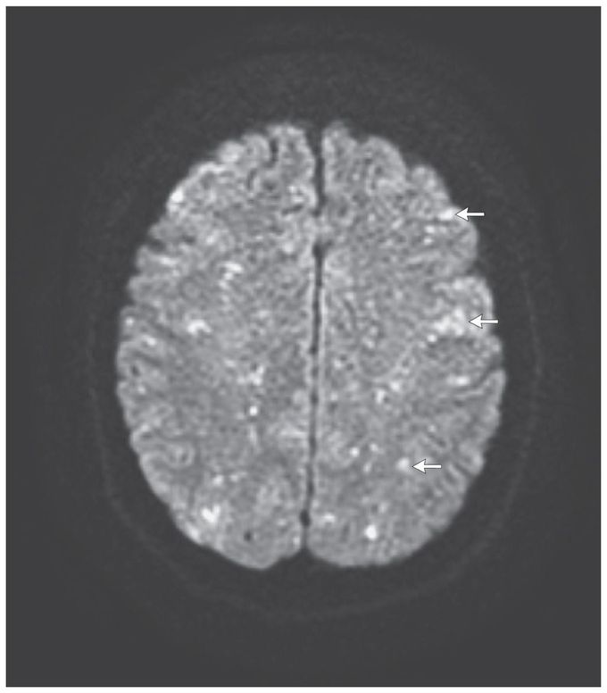


Cerebral Fat Embolism
A 56-year-old office worker underwent elective hip replacement after a right-hip fracture. She remained somnolent a few hours after surgery and required intubation. She had roving eye movements and decerebrate posturing. Two petechiae were evident on the left arm and a single petechia in the right axilla. Magnetic resonance imaging (MRI) of the brain showed numerous pinpoint, hyperintense foci in both the gray and the white matter of the cerebral and cerebellar hemispheres, the thalami, and the caudate heads bilaterally on T2-weighted images. On diffusion-weighted images, there were innumerable punctate foci of restricted diffusion, producing a “starfield” appearance (arrows). The MRI findings in this context were consistent with the cerebral fat embolism syndrome. The hyperintensities noted on diffusion-weighted images have been hypothesized to represent foci of cytotoxic edema due to acute cerebral microinfarcts, whereas hyperintensities on T2-weighted images presumably reflect vasogenic edema. The majority of patients with systemic fat embolism have reversible neurologic deficits. Our patient's neurologic recovery was slow, but about 4 months after the injury, she reported being essentially back to baseline, her only residual deficits being mild difficulty with word retrieval and short-term memory.
I love love your content, they're so very informative!!!

