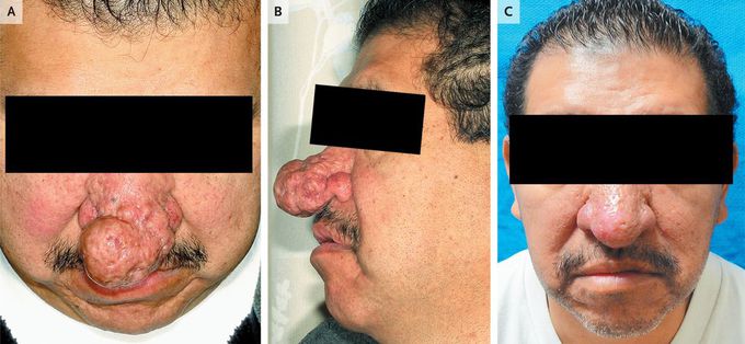


Rhinophyma
A 42-year-old man with a 10-year history of rosacea presented with impaired nasal breathing and a mass on the tip of his nose that began growing 9 months earlier. Examination revealed a multilobulated sebaceous nodule (4 cm by 3 cm) protruding from the nasal tip (Panels A and B). The histopathological findings of marked sebaceous hyperplasia, follicular rupture, an absence of granulomas, and prominent fibrosis confirmed the clinical suspicion of rhinophyma. A biopsy specimen was obtained, and staining did not reveal infectious organisms. Phyma is the result of hyperplasia and fibrosis of the sebaceous glands in the presence of rosacea. Although rhinophyma is by far the most common pattern in cases of phyma, metophyma (swelling of the forehead), otophyma (swelling of the ear), and gnathophyma (swelling of the chin) can also be observed. The lesions can become large, causing significant social stigmatization and posing a challenge in the management of patient care. Recontouring with the use of electrosurgery or CO2 laser resurfacing is common. This patient underwent staged procedures, with shave–debulking surgery followed by contouring with electrosurgery (Panel C). His breathing was restored to normal.


