


Orbital Hydatid Cyst
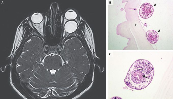
A 31-year-old woman presented to the emergency department with a history of blurred vision in the left eye over a 4-week period and progressive bulging of the left eye over a 2-week period. Physical examination revealed nontender proptosis of the left eye, paresis of the left abducens nerve, and reduced visual acuity in the left eye. A T2-weighted gadolinium-enhanced magnetic resonance image of the brain showed a well-defined, ovoid, cystic, and retrobulbar lesion in the left orbital cavity (Panel A). The optic nerve was displaced nasally and the lateral rectus muscle was compressed. The patient underwent left lateral orbitotomy, and the cyst was completely removed but ruptured during surgery. The area was washed with saline. Histopathological examination revealed multiple protoscolices (Panel B, arrowheads), with central hooklets (Panel C, arrow), adjacent or attached to a thick, acellular laminated echinococcal cyst membrane (Panel B, asterisk). A diagnosis of a hydatid cyst caused by the Echinococcus granulosus tapeworm was made. Thoracic and abdominal computed tomographic scans revealed no extraorbital organ involvement. The patient received a 3-month course of albendazole, and at follow-up 3 months later, she had full recovery of visual acuity.

So if there is cyst formation it would be causes of foreign bodies??or any other way?
