


Abdominal Ectopic Pregnancy
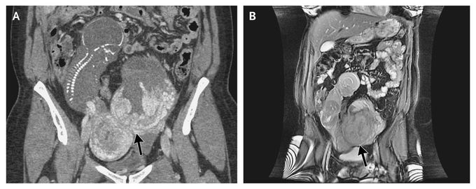
A 30-year-old woman who had a history of two pregnancies and one birth presented with an uncomplicated pregnancy until routine ultrasonography at 19 weeks revealed severe oligohydramnios and a fetus that appeared to be extrauterine. Computed tomography (Panel A) and magnetic resonance imaging (Panel B) of the abdomen and pelvis confirmed an abdominal ectopic pregnancy, with no uterine wall visible surrounding the pregnancy. The fetus was visualized in the right abdomen with a crown-to-rump length of 14 cm, with the placenta attached to the serosa of the uterine fundus (Panel A, arrow). No amniotic fluid surrounded the fetus. The pregnancy was terminated, and surgical removal of the fetus was performed. An abdominal pregnancy refers to a pregnancy that has implanted in the peritoneal cavity, external to the uterine cavity and fallopian tubes. In contrast to tubal ectopic pregnancies, abdominal pregnancies may go undetected until an advanced gestational age. Abdominal pregnancies are associated with a high rate of maternal complications.
It says the pregnancy was terminated. Can believe her pregnancy went 19 weeks!!
https://www.facebook.com/PrimalTRTOfficial/Women’s Reproductive Health & Daily WellnessFlexiLeafhttps://www.facebook.com/HumeHealthBodyPodOfficial/Pulsar Vexline Review 2026 - Legit Or Scam Trading Platform? Fact CheckGelatine Sculpt | Official WebsiteBurnSlim Official | Support Your Weight Management Journey
