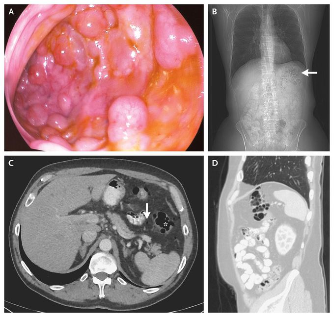


Pneumatosis Intestinalis
A 63-year-old man presented for a screening colonoscopy. During the colonoscopy, several submucosal polypoid lesions, 3 to 8 mm in diameter, were noted in the splenic flexure. Since the lesions were easily indented with gentle pressure and had a bluish hue, pneumatosis intestinalis was suspected (Panel A). When the lesions were punctured, they became deflated, supporting the diagnosis (see video). Computed tomography revealed multiple air pockets in the intestinal wall at the splenic flexure (Panel B, arrow; Panel C, asterisk; and Panel D) and some free intraperitoneal air (Panel C, arrow). The results of histopathological inspection of an endoscopic-biopsy specimen were consistent with pneumatosis intestinalis. Pneumatosis intestinalis is diagnosed by the presence of air pockets in the intestinal wall. In certain cases, pneumatosis intestinalis may be considered a surrogate marker for intestinal ischemia and impending perforation. However, the condition may also occur in a benign context and is no longer considered a disease but rather a sign, and its significance needs to be considered in accordance with each patient's clinical context. This patient required no specific treatment and has remained asymptomatic.

