


Hampton's Hump
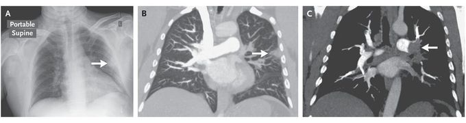
A 46-year-old man presented to the emergency department with the sudden onset of dyspnea, pleuritic chest pain, and hypoxemia. The patient's medical history and family history were unremarkable. An electrocardiogram showed a deep S wave in lead I, a large Q wave in lead III, an inverted T wave in lead III, and an incomplete right bundle-branch block, suggesting right ventricular strain. The patient underwent chest radiography, which revealed a Hampton's hump on the left side of the chest (Panel A, arrow). Computed tomographic pulmonary angiography further defined this area of pulmonary infarction (Panel B, arrow) and revealed bilateral pulmonary emboli, including a thrombus in the left main pulmonary artery (Panel C, arrow). Originally described in 1940 by Hampton and Castleman, Hampton's hump is a peripheral wedge-shaped opacification abutting the pleura, signifying pulmonary infarction distal to a pulmonary embolism. The majority of pulmonary emboli do not result in infarction of the distal lung, presumably because of vascular collateralization. Infarction is most likely to occur in patients with large pulmonary emboli or underlying lung disease. Anticoagulation therapy was initiated, and the patient was discharged home in stable condition.
