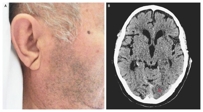


Bilateral Earlobe Creases
An 84-year-old man with hypertension, diabetes, and hypercholesterolemia presented to the emergency department with a 6-hour history of visual difficulty. Physical examination revealed a right homonymous hemianopia and no other relevant neurologic findings. A diagonal crease in each earlobe (Frank's sign) was noted (Panel A). Urgent computed tomography revealed a subacute occipital infarction in the territory of the left posterior cerebral artery (Panel B, asterisk), as well as many other old ischemic lesions. Frank's sign was originally described as a marker of coronary artery disease, with a moderate sensitivity (approximately 48%) and specificity (approximately 88%). This sign has been subsequently associated with other cardiovascular risk factors. The patient was treated conservatively, his course was uneventful, and he was discharged home 1 week after presentation, with persistence of the visual deficit.


