


An Unknowingly Swallowed Inedible Toy
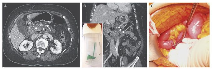
A 72-year-old woman presented to the emergency department with a 3-day history of periumbilical pain. She had had one episode of vomiting. She had no other notable medical history. Physical examination showed a temperature of 38°C and right abdominal tenderness without contracture. Laboratory tests showed a C-reactive protein level of 256 mg per liter and a white-cell count of 19,300 per cubic millimeter, with 90% neutrophils. A contrast-enhanced computed tomographic (CT) scan of the abdomen obtained during the portal venous phase shows infiltration of the root of the mesentery surrounding the superior mesenteric artery in association with pneumoretroperitoneum (Panel A, arrows). A CT reconstruction image in the coronal plane shows hypoattenuation of an ax-shaped foreign body perforating the third portion of the duodenum (Panel B, arrows). An exploratory laparotomy revealed the cause of the perforation (Panel C, green plastic gripped by the forceps), which was a plastic ax that was 2 cm long (Panel B inset). The patient reported that she had eaten a Christmas cake 7 days earlier. The perforation was repaired with sutures, and the patient recovered and was discharged after 10 days of hospitalization.
