


Roth Spots in Infective Endocarditis
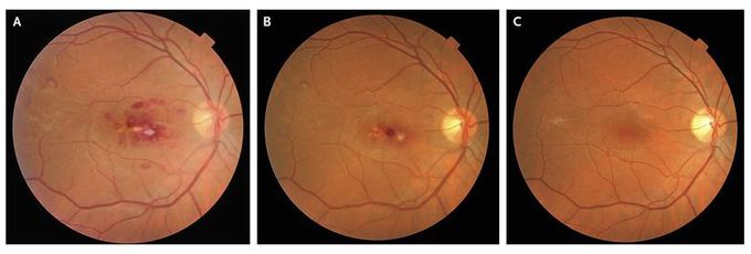
A 34-year-old man presented to the eye emergency department with reduced vision in the right eye that had developed that morning. He was well, apart from episodes of dizziness and dyspnea after exercise during the previous 4 months. These episodes had started 1 week after the patient had undergone a dental treatment. Previous extensive investigations had been inconclusive. His retinal appearance and symptoms prompted referral to the cardiology team, which admitted him that day. He was afebrile but had a pansystolic murmur and a solitary splinter hemorrhage on the right thumb. Echocardiography revealed moderate-to-severe mitral-valve regurgitation, with thickening and signs of vegetations. The ejection fraction was 65%. The other values were normal. Four sets of blood cultures yielded Streptococcus viridans, and intravenous antibiotics were started immediately. His symptoms improved, and his visual acuity gradually improved from 20/200 at presentation to 20/20 8 months after presentation. His right fundal appearance is shown, at presentation (Panel A), 3 days later (Panel B), and 3 months later (Panel C). The presence of white-centered hemorrhages (Roth spots) should prompt the consideration of possible infective endocarditis. A comparison of Panel A and Panel B (3 days apart) shows how quickly such spots can change. Monitoring of the patient's mitral-valve regurgitation, which was started after complete resolution of the endocarditis, is ongoing.
