


Bilateral Wandering Lens in an Infant
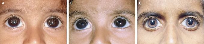
A healthy 1-year-old boy was brought to the emergency department by his parents, who had observed the sudden development of a globulelike mass in both of his eyes. He did not seem to have any obvious visual difficulty. On examination, it was noted that the spheroidal transparent crystalline lens in both eyes had dislocated into the anterior chamber (Panel A). When the patient lay supine for ultrasonography (Panel B), the lenses spontaneously returned to the vitreous cavity (Panel B), without any pharmacologic dilation of the pupils. The migration of the lenses may have been due to a squeezing action of the lids and the pressure of the ultrasound probe. The results of physical examination were otherwise normal. Examination of the eyes with the patient under anesthesia revealed normal corneal clarity, with a corneal diameter of 13 mm (reference range, 11 to 12) and an intraocular pressure of 14 mm Hg in both eyes. The crystalline lens in each eye was freely floating in the vitreous cavity. The patient underwent sequential pars plana lensectomy and vitrectomy. Bilateral aphakic glaucoma developed over the next 5 years, and intraocular pressure was controlled by means of two antiglaucoma medications, a prostaglandin analogue and a topical carbonic anhydrase inhibitor. At the most recent follow-up visit, 10 years after the initial presentation (Panel C), the visual acuities were 20/25 and 20/50 and the intraocular pressures were 12 mm Hg and 13 mm Hg in the right and left eyes, respectively. Lifelong periodic follow-up will continue in order to control the patient's intraocular pressure and monitor visual function.
