


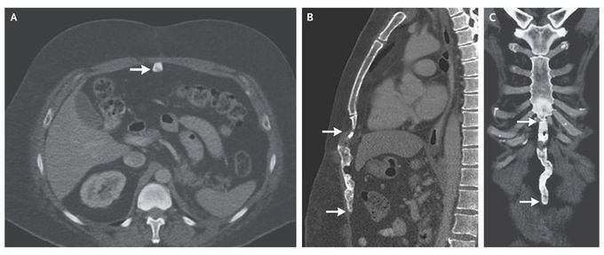
Heterotopic Ossification of a Midline Abdominal Incision
A 49-year-old man underwent computed tomography (CT) as part of routine follow-up after a left nephrectomy performed 2 years previously for renal-cell carcinoma. A healed vertical midline incision in the upper abdomen was found during a physical examination. CT of the abdomen revealed a vertically oriented, linear, calcified lesion in the incision scar on the anterior abdominal wall (Panel A, arrow), extending from the immediate subxiphoid region (Panels B and C, top arrow) to the umbilical region (Panels B and C, bottom arrow). This finding was consistent with heterotopic ossification, a subtype of myositis ossificans traumatica. Histologic evidence of osseous, cartilaginous, and occasionally myelogenous elements distinguishes this entity from dystrophic calcification. Although it is symptomatic only in rare cases, it may cause abdominal pain and discomfort; treatment in such cases consists of complete excision with primary closure. In this case, no further action was taken; however, the patient was advised to return for a follow-up visit.
