


Morphologic Changes in Erythrocytes in Hypertriglyceridemia
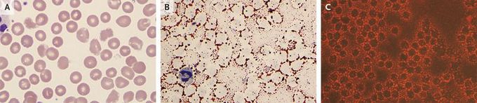
A 39-year-old woman with a history of gestational diabetes was admitted with epigastric pain from acute pancreatitis. Ultrasonography showed no gallstones or bile-duct dilatation. She had no history of hyperlipidemia, but multiple blood samples were grossly lipemic, and serum triglyceride levels were markedly increased at more than 4425 mg per deciliter (50.0 mmol per liter), as compared with a normal value of less than 200 mg per deciliter (2.3 mmol per liter). Wright's staining of a peripheral-blood smear showed numerous small lipid droplets overlying and surrounding the periphery of the red cells (Panel A). Staining with oil red O, which is commonly used to demonstrate lipid in tissue sections, was performed on the peripheral blood and highlighted the triglycerides surrounding the red cells on light microscopy (Panel B) and under fluorescence (Panel C). The patient underwent treatment for severe hypertriglyceridemia and pancreatitis. The triglyceride level decreased to 471 mg per deciliter (5.3 mmol per liter), and the peripheral blood showed a marked decrease in staining with oil red O on light microscopy and under fluorescence. The symptoms of pancreatitis resolved with treatment, and the patient was discharged home.
