


Destructive Ulcerated Lesions of the Hard Palate
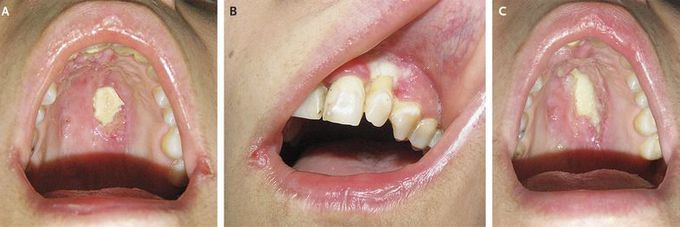
A 34-year-old woman presented with a 3-month history of ulcerated plaques on the palate and gingiva. Physical examination revealed two ulcerated lesions with irregular margins on the hard palate (Panel A) and gingival mucosa (Panel B). The palatal bone and maxillary alveolar process were focally denuded of their mucosal covering. Computed tomography of the paranasal sinuses, nose, and orbit with the use of contrast material revealed no evidence of mass or lymphadenopathy. Nasal endoscopy was normal. Culture of a palatal biopsy specimen was positive for Staphylococcus aureus. Histopathological findings were negative for cancer. The patient began receiving intravenous antibiotic agents and was doing well until day 10, when she presented with fever and intermittent swelling on the left side of her face. Two weeks later, the palatal lesion had enlarged (Panel C), and a second biopsy was performed. Histopathological examination revealed an extranodal natural-killer-cell–T-cell lymphoma, nasal type. This is a non-Hodgkin's lymphoma with extranodal presentation within the upper aerodigestive tract, commonly involving the nasal cavity, nasopharynx, and paranasal sinuses. The patient received radiotherapy and modified SMILE chemotherapy (consisting of asparaginase, methotrexate, ifosfamide, etoposide, and dexamethasone) and underwent autologous bone marrow transplantation. At 8 months, she remains in remission, with no evidence that the lymphoma has recurred.
