


Mobile Large Left Atrial Thrombus
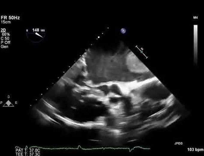
A 58-year-old obese woman with rheumatic heart disease who had undergone balloon mitral valvuloplasty 13 years earlier presented with progressively increasing dyspnea on exertion for the previous month. She had no history of systemic embolization and was not receiving anticoagulation. The physical examination revealed atrial fibrillation with a ventricular rate of 90 beats per minute. The findings on cardiovascular examination were consistent with severe mitral stenosis and pulmonary hypertension (right parasternal heave, variable but loud first heart sound, loud second heart sound, and mid-diastolic murmur at apex). Transesophageal echocardiographic examination confirmed severe rheumatic mitral stenosis (Panel A, arrow) with severe pulmonary hypertension (estimated mitral orifice size, 0.8 cm2; pulmonary-artery systolic pressure, 85 mm Hg). Also visible was a 4.5-cm left atrial mass, which was thought likely to be a thrombus, freely floating within the left atrial cavity (Panel B). The mass intermittently dropped into the mitral orifice, temporarily occluding it, before bouncing away again (video). The large size of the mass and the presence of severe mitral stenosis prevented it from migrating into the systemic circulation with catastrophic consequences. The patient underwent surgery, which confirmed the mass to be a large thrombus. Open mitral commissurotomy was performed along with removal of the thrombus, and the patient had an uneventful recovery.
