


An Unusual Tophus
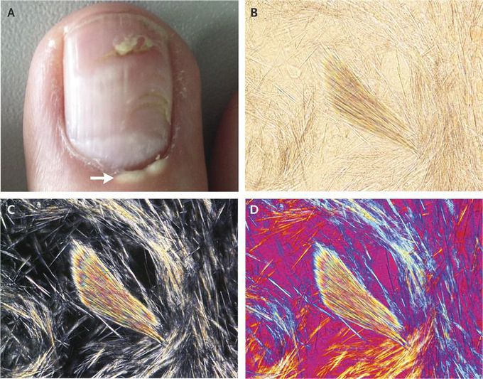
A 77-year-old man presented for evaluation of a painless white periungual lesion in the cuticle that he had noted 2 days earlier (Panel A, arrow). The patient had received a diagnosis of gout the preceding week, when he presented with acute inflammatory changes in his left wrist and analysis of aspirated synovial fluid showed urate crystals. In addition, tophi were seen on both elbows. Prednisone and colchicine were prescribed. On the subsequent visit, the white appearance of the periungual finding raised the question of another focus of gout. Microscopy of a sample obtained from the cuticle showed abundant monosodium urate crystals in ordinary light (Panel B), polarized light (Panel C), and compensated polarized light (Panel D). The cuticle lesion probably damaged the ungual matrix, resulting in alterations of the nail. After 4 months of allopurinol treatment, the periungual deposits had disappeared and the remaining tophi had diminished in size. Tophaceous deposits may occur in a variety of atypical places, including around a fingernail or toenail, and should be considered as potential causes of unusual lesions in a patient with a history of gout.
