


Peritoneal Loose Body
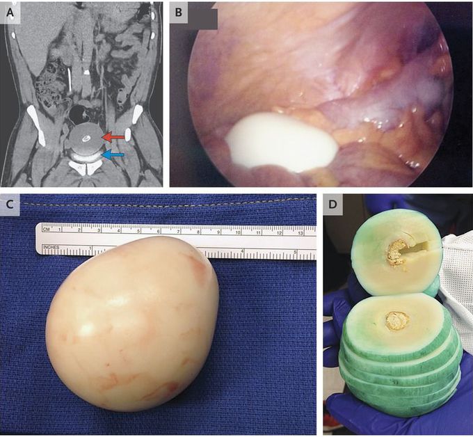
A 62-year-old man presented for evaluation of a history of urinary frequency of more than 20 years. The physical examination and laboratory findings were unremarkable. Computed tomography of the abdomen and pelvis revealed an 8.5-cm midline mass with central calcification (Panel A, red arrow) superior to and compressing the bladder (Panel A, blue arrow). Laparoscopy revealed a free-floating, smooth, firm, rubbery mass measuring 10 cm by 9.5 cm by 7.5 cm and weighing 220 g (Panels B and C). The sectioned specimen included several layers; green ink was used to delineate section margins (Panel D). Histologically, the mass contained predominantly acellular, laminated, fibrous tissue; centrally, the specimen contained proteinaceous material with fibrinoid necrosis, surrounded by a ring of calcification. The findings were consistent with a peritoneal loose body, a formation that is thought to result from torsed, infarcted, and detached epiploic appendages that transform into fibrotic masses. Such masses are often asymptomatic when they are small, but they can be large enough to cause extrinsic compression that is associated with bowel obstruction, urinary retention, or (as in this patient) urinary frequency. The patient's urinary frequency resolved immediately after the surgical removal of the mass.
Lol same. But then again how do you get an egg in your abdomen without perforating anything.
