


Central Retinal-Vein Occlusion
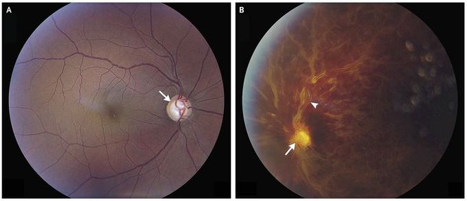
A 43-year-old man with hypertension presented with sudden painless loss of vision in his left eye. Visual acuity was measurable only by ability to count fingers at a distance of 3 feet. A relative afferent pupillary defect was also noted. Intraocular pressure was at the upper limit of the normal range, at 20 mm Hg bilaterally. Funduscopic examination was remarkable for optic-nerve cupping of the right eye (Panel A, arrow) and hyperemia and swelling of the optic nerve (Panel B, arrow), macular edema, diffuse intraretinal hemorrhages, and dilated and tortuous retinal veins (Panel B, arrowhead) in the left eye. A diagnosis of central retinal-vein occlusion of the left eye and bilateral open-angle glaucoma was made. The differential diagnosis of sudden painless vision loss includes vitreous hemorrhage, retinal artery or vein occlusion, retinal detachment, ischemic optic neuropathy, and occipital stroke. Central retinal-vein occlusion is an important and often dramatic cause of sudden painless loss of vision. Risk factors include older age, hypertension, glaucoma, and diabetes mellitus. Inflammatory causes and coagulopathy should be considered in younger patients. The patient was treated with timolol ophthalmic drops and antihypertensive medications. He received serial intravitreal injections of bevacizumab in the left eye, with improvement of visual acuity to 20/200 and decreased macular edema over an 8-month period of follow-up.
