


Female reproductive disorders
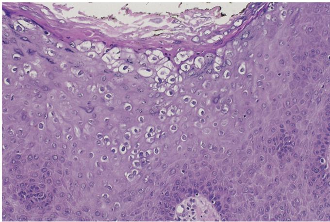
A 36-year-old sexually active woman has noticed that warty vulvar lesions have been increasing in size and number over the past 5 years. On physical examination, there are multiple 0.5- to 2-cm, red-pink, flattened lesions with rough surfaces present on the vulva and perineum. One of the larger lesions is excised; its microscopic appearance is shown in the figure. Which of the following infectious agents is most likely to produce these lesions? A. Candida albicans B. Chlamydia trachomatis C. Haemophilus ducreyi D. Human papillomavirus E. Treponema pallidum The epithelium shows typical features of infection with human papillomavirus (HPV)—specifically, prominent perinuclear vacuolization (koilocytosis) and angulation of nuclei. These lesions, called condylomata acuminata, may occur anywhere on the anogenital surface, as single lesions or, more commonly, as multiple lesions. They are not precancerous. Condylomata are associated with HPV infection, often types 6 and 11.
