


Lancisi’s Sign
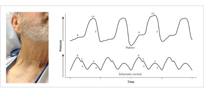
A 60-year-old man with nonischemic cardiomyopathy presented with progressive dyspnea and weight gain of approximately 9 kg that had developed over a period of 2 to 3 weeks. On examination, a grade 2/6 holosystolic murmur that augmented with inspiration was noted at the left lower sternal border. Examination of the neck revealed a palpable, monomorphic venous pulsation, known as Lancisi’s sign (see video). Transthoracic echocardiography revealed malcoaptation of the tricuspid-valve leaflets as a result of annular dilatation, with resultant severe regurgitation. Lancisi’s sign is a physical finding of severe tricuspid regurgitation. Normally, three peaks and two troughs characterize the venous waveform (the lower strip is a generic representation of normal findings, for comparison). The first peak, called the a wave, results from atrial contraction during late diastole. Next, during early systole, isovolumetric ventricular contraction triggers closure of the tricuspid valve, producing the c wave. In mid-systole, given a competent tricuspid valve, a combination of atrial relaxation and descent of the atrial floor during ventricular contraction results in the x descent. The third peak, the v wave, occurs as a result of atrial filling during late systole. Finally, passive ventricular filling in early diastole produces the y descent. In the context of tricuspid regurgitation, retrograde blood flow into the right atrium during ventricular systole results in loss of the x descent (the upper strip shows the right atrial pressure tracing from this patient), creating a fused cv wave that appears as a large pulsation within the internal jugular vein that is often palpable. This wave is typically followed by an augmented y descent, which is the consequence of an increased pressure gradient between the right atrium and right ventricle. In this patient, diuretic agents were used to normalize the volume status, and the symptoms abated.

In nonischaemic heart failure, what happens most commonly is dilation of the heart's main pump(left ventricle), in a condition called dilated cardiomyopathy. Initially, to compensate for the heart's pump insufficiency in pumping blood, the ventricle dilates and enlarges to compensate for the reduced pumping activity, as such, the blood output is maintained. Eventually, the heart's pump loses the strength to do so and hence pumps even lesser amount of blood than required. In response, the right ventricle soon joins; due to increased retention in the left ventricle, the pressure increases thereby decreasing pulmonary venous return and a corresponding rise in pressure which soon reaches the right ventricle. The increase in the right ventricular pressure decreases systemic venous return/emptying. Progressively, the heart retains more blood and then the kidneys retain more fluid and edema develops, commonly starting from both of the lower extremities and later involving the entire body. The weight gain that occurs is most likely hence attributable to the generalised oedema. From the little I know.🙂
