


Scalp Necrosis Associated with Giant-Cell Arteritis
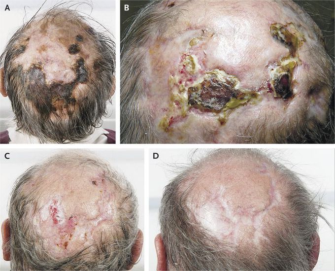
A 74-year-old man presented with a 3-month history of headache, jaw claudication, and worsening scalp ulceration. He reported no visual loss or polymyalgia rheumatica. Examination revealed areas of scalp necrosis (Panel A). Both temporal arteries were difficult to palpate but were nontender. His erythrocyte sedimentation rate was 64 mm per hour, and his C-reactive protein level 45 mg per liter. He received a diagnosis of scalp necrosis as a complication of giant-cell arteritis. Treatment was initiated with glucocorticoids (60 mg of prednisolone per day) and topical dressings that were administered daily. During the next few months, his symptoms decreased and the scalp lesions started to heal (Panels B and C). The dose of prednisolone was tapered by 10 mg every month until it reached 20 mg per day. The dose was then tapered more slowly until the lesions healed completely (Panel D), after which the medication was discontinued. Scalp necrosis is a rare and potentially life-threatening complication of giant-cell arteritis. Early recognition and treatment are important.
