


Heliaover 6 years ago
Radiological findings of osteoarthritis
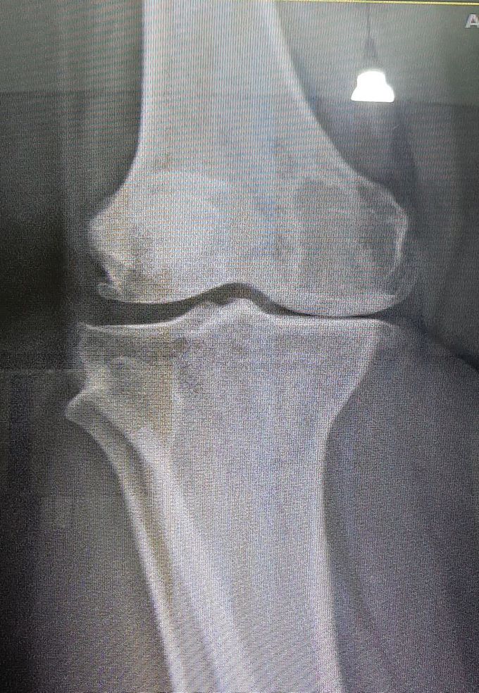
Here's a radiography of a 63-year's-old woman whose chief complaint is knee pain which worsens during walking. Tell the four major radiologic findings, consistent with osteoarthritis. And also, anyone knows the Kellgren's classification of osteoarthritis? Let's review what we know!
Top rated comment
over 6 years ago
One is: the distal part of the phemur is in direct contact with the proximal part of the tibia. I'm trying to use some terminology, I'm in my second week of radiology. So please be gentle ☺
Other commentsSign in to post comments. You don't have an account? Sign up now!
over 6 years ago
tibial plate sclerosis, flattening of femoral condyles, osteophytes, reduced joint space, cysts in medial condyle
