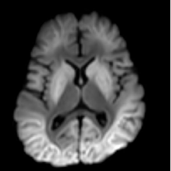

A 3-year-old boy presents with fever, seizures, and other clinical features suggestive of encephalopathy Acute leukoencephalopathy with restricted diffusion (ALERD) Background: Acute leukoencephalopathy with restricted diffusion is a clinical-radiological spectrum characterized by the clinical features of acute encephalopathy and neuroimaging showing diffusion restriction predominantly in the subcortical white matter. Etiologically, it is classified as Infectious ALERD or Toxic ALERD, based on whether it occurs in the setting of infection or toxins, respectively. Clinical Presentation: ALERD presents as acute encephalopathy. Imaging features may not be evident in first 2 to 3 days. They appear at the third to ninth days after symptom onset. Key Diagnostic Features: There are two distinct imaging patterns recognized: diffuse type and central sparing type. In the diffuse type, diffusion restriction is seen involving bilateral subcortical white matter. In the central sparing type, there is sparing of central regions of the brain, mainly the primary sensory motor cortex. The diffusion restriction in subcortical white matter appears similar to multiple dendritic bright tree branches, which is characteristically described as "bright tree appearance." Differential Diagnosis: Atypical PRES and ADEM: Changes are more pronounced on T2/FLAIR than DWI Postictal changes: T2/FLAIR hyperintensities are usually seen in both the cortex and subcortical white matter. Clinical symptoms are transient. Febrile infection-related epilepsy syndrome (FIRES): changes are seen in gray matter structures such as the mesial temporal lobes, basal ganglia, and/or hippocampi Mild leukoencephalopathy with reversible splenial lesion (MERS): Lesions are mainly restricted to the splenium of corpus callosum

